Home › Unlabelled › Lower Leg Bone Diagram - The Lower Limbs Human Anatomy And Physiology Lab Bsb 141
Lower Leg Bone Diagram - The Lower Limbs Human Anatomy And Physiology Lab Bsb 141
Lower Leg Bone Diagram - The Lower Limbs Human Anatomy And Physiology Lab Bsb 141. In addition, the broad hip bones provide protection to the delicate internal organs of the pelvis, such as the intestines, urinary bladder, and uterus. Bone diagram forehead (frontal bone) nose bones (nasals) cheek bone (zygoma) upper jaw (maxilla) lower jaw (mandible) breast bone (sternum) upper arm bone (humerus) lower arm bone (ulna) thigh bone (femur) collar bone (clavicle) toe bones (phalanges) ankle bones (tarsals) kneecap (patella) shin bone (tibia) calf bone (fibula) foot bones The lower leg is made up of two very strong, long bone—the tibia and the fibula. These bones may break into two or more pieces.if a broken bone has been exposed to the outside, either by a cut over the fracture, or by bone sticking out through the skin, it is called an open fracture. Some types of leg pain can be traced to problems in your lower spine.
The fibula or calf bone is a leg bone on the lateral side of the tibia, to which it is connected above and below.it is the smaller of the two bones and, in proportion to its length, the slenderest of all the long bones. It is located toward the middle of the lower leg. Anatomical structures of the lower limb (hip, thigh, knee, leg, ankle and foot) and specific regions (compartment of the lower limb) are visible on dynamic labeled images. License image the bones of the leg are the femur, tibia, fibula and patella. These bones may break into two or more pieces.if a broken bone has been exposed to the outside, either by a cut over the fracture, or by bone sticking out through the skin, it is called an open fracture.
Arrangement Of Lower Limb Bones from wodeshu.gitee.io It lies within the quadriceps tendon. Our goal is that these leg anatomy worksheets pictures gallery can be a direction for you, bring you more references and also make you have a great day. This large tendon from the powerful thigh muscles (quadriceps) wraps round the patella and is attached to the top of the lower leg bone (tibia). In addition, the broad hip bones provide protection to the delicate internal organs of the pelvis, such as the intestines, urinary bladder, and uterus. Many muscles that move the trunk and legs, such as our abdominal muscles, attach to the hip bones. It is located toward the middle of the lower leg. While their parts are similar in general, their structure has been adapted to differing functions. In this small section, we'll briefly mention the main parts of the leg, namely the bones, muscles, and neurovasculature.
The head of the femur forms the ball and socket hip joint with the acetabulum of the hip bone.
Bone diagram forehead (frontal bone) nose bones (nasals) cheek bone (zygoma) upper jaw (maxilla) lower jaw (mandible) breast bone (sternum) upper arm bone (humerus) lower arm bone (ulna) thigh bone (femur) collar bone (clavicle) toe bones (phalanges) ankle bones (tarsals) kneecap (patella) shin bone (tibia) calf bone (fibula) foot bones The proximal portion of the tibia is tibial plateau which acts as a cusp for the knee, the distal portion tapers into the medial malleoli and the concave surface which articulates with the talus at the ankle joint. At the distal end of the femur, two rounded condyles meet the tibia and fibula bones of the lower leg to form the knee joint. Also called the shin bone, the tibia is the longer of the two bones in the. Blood and bones kids drawings. Ankle and foot bones and joints unit 4/12/18 lower leg: The patella is the kneecap bone. Out of these, the cookies that are categorized as necessary are stored on your browser as they are essential for the working of basic functionalities of the website. These bones may break into two or more pieces.if a broken bone has been exposed to the outside, either by a cut over the fracture, or by bone sticking out through the skin, it is called an open fracture. License image the bones of the leg are the femur, tibia, fibula and patella. While their parts are similar in general, their structure has been adapted to differing functions. Most leg pain results from wear and tear, overuse, or injuries in joints or bones or in muscles, ligaments, tendons or other soft tissues. Some types of leg pain can be traced to problems in your lower spine.
Leg femur diagram data wiring diagram today. It is located toward the middle of the lower leg. The human leg, in the general word sense, is the entire lower limb of the human body, including the foot, thigh and even the hip or gluteal region. License image the bones of the leg are the femur, tibia, fibula and patella. Also called the shin bone, the tibia is the longer of the two bones in the.
Anatomy Of The Lower Limb 18 Download Scientific Diagram from www.researchgate.net The lower extremity, commonly referred to as the leg, contains four bones (the femur, the patella, the tibia, and the fibula) and bends at the hip, the knee, and the ankle. Bone diagram forehead (frontal bone) nose bones (nasals) cheek bone (zygoma) upper jaw (maxilla) lower jaw (mandible) breast bone (sternum) upper arm bone (humerus) lower arm bone (ulna) thigh bone (femur) collar bone (clavicle) toe bones (phalanges) ankle bones (tarsals) kneecap (patella) shin bone (tibia) calf bone (fibula) foot bones These bones may break into two or more pieces.if a broken bone has been exposed to the outside, either by a cut over the fracture, or by bone sticking out through the skin, it is called an open fracture. The tibia (also called the shinbone) is located near the midline of the leg. The head of the femur forms the ball and socket hip joint with the acetabulum of the hip bone. The human leg, in the general word sense, is the entire lower limb of the human body, including the foot, thigh and even the hip or gluteal region. The tibia, also known as the shin bone, is the stronger and larger of the two. Ankle and foot bones and joints unit 4/12/18 lower leg:
Some types of leg pain can be traced to problems in your lower spine.
In the realm of anatomy, the 'leg' is strictly the region between the knee and the ankle joints rather than the entire lower extremity, as erroneously referred to in common language. Leg pain can also be caused by blood clots, varicose veins or poor circulation. Legs are used for standing, and all forms of. The hip joint gives the leg an incredible range of motion while still providing support to the body's weight. This website uses cookies to improve your experience while you navigate through the website. The lower extremity, commonly referred to as the leg, contains four bones (the femur, the patella, the tibia, and the fibula) and bends at the hip, the knee, and the ankle. The patella is the kneecap bone. Related posts of muscles and tendons of the leg muscle anatomy in shoulder. The medial side of the tibia is located immediately under the skin, allowing it to be easily palpated down the entire length of the medial leg. At the distal end of the femur, two rounded condyles meet the tibia and fibula bones of the lower leg to form the knee joint. He leg's main function in the human is for locomotion and support of the rest of the body. While factors like what your pain feels like—stabbing, burning, or cramping, and so on—can provide insight, oftentimes, a detailed physical examination and/or an imaging test are needed to clinch the diagnosis. The tibia (also called the shinbone) is located near the midline of the leg.
The lower leg is made up of two very strong, long bone—the tibia and the fibula. Learn vocabulary, terms, and more with flashcards, games, and other study tools. Diagram and names of leg bones, diagram of foot and leg bones, diagram of leg bones, diagram of lower leg bones, diagram of the bones in your leg, bone, diagram and. The quadriceps muscles straighten the knee. Our goal is that these leg anatomy worksheets pictures gallery can be a direction for you, bring you more references and also make you have a great day.
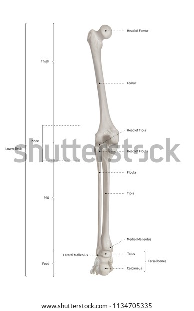
Infographic Diagram Human Skeleton Lower Limb Stock Illustration 1134705335 from image.shutterstock.com This large tendon from the powerful thigh muscles (quadriceps) wraps round the patella and is attached to the top of the lower leg bone (tibia). The knee joint is the largest joint in the body and is primarily a hinge joint, although some sliding and rotation occur. Ankle and foot bones and joints unit 4/12/18 lower leg: Leg pain can also be caused by blood clots, varicose veins or poor circulation. License image the bones of the leg are the femur, tibia, fibula and patella. The lower leg contains two major long bones, the tibia and the fibula, which are both very strong skeletal structures. This area is commonly referred to as the calf. At the distal end of the femur, two rounded condyles meet the tibia and fibula bones of the lower leg to form the knee joint.
Leg femur diagram data wiring diagram today.
Includes labels for muscles, bones, nerves and arteries of the knee, leg and foot. In the realm of anatomy, the 'leg' is strictly the region between the knee and the ankle joints rather than the entire lower extremity, as erroneously referred to in common language. This large tendon from the powerful thigh muscles (quadriceps) wraps round the patella and is attached to the top of the lower leg bone (tibia). The lower leg is comprised of two bones, the tibia and the smaller fibula. The quadriceps muscles straighten the knee. The proximal portion of the tibia is tibial plateau which acts as a cusp for the knee, the distal portion tapers into the medial malleoli and the concave surface which articulates with the talus at the ankle joint. While their parts are similar in general, their structure has been adapted to differing functions. The smaller lateral bone of the lower leg. Also called the shin bone, the tibia is the longer of the two bones in the. It is located toward the middle of the lower leg. Muscle anatomy in shoulder 12 photos of the muscle anatomy in shoulder muscle anatomy neck and shoulder, muscle anatomy of shoulder, muscle anatomy of shoulder joint, muscle anatomy shoulder back, muscle anatomy shoulder upper arm, human muscles, muscle anatomy neck and shoulder, muscle anatomy of shoulder, muscle. This area is commonly referred to as the calf. Anatomical structures of the lower limb (hip, thigh, knee, leg, ankle and foot) and specific regions (compartment of the lower limb) are visible on dynamic labeled images.
These bones may break into two or more piecesif a broken bone has been exposed to the outside, either by a cut over the fracture, or by bone sticking out through the skin, it is called an open fracture leg bone diagram. Also called the shin bone, the tibia is the longer of the two bones in the.


comment 0 comments
more_vert